A specialized retina specialist focuses on complex medical conditions affecting the macula and the neural tissue at the back of the eye. These medical professionals complete specialized training, and they gain experience through extensive fellowship programs to master advanced surgical techniques. Because untreated retinal disorders can cause vision loss without intervention, physicians rely on these experts to evaluate abnormalities in the eye and its surrounding structures.
Testing
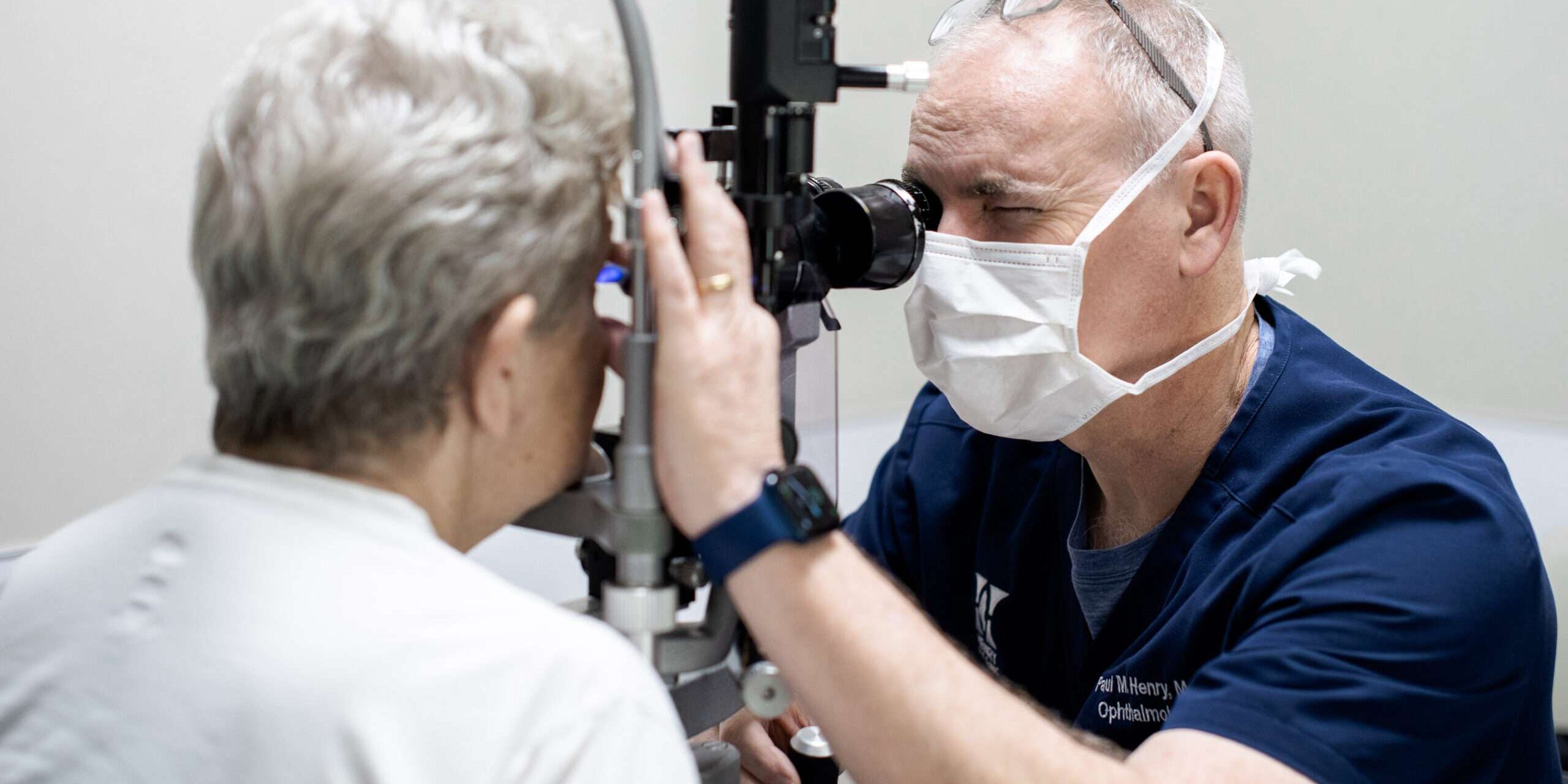
A retina specialist often relies on diagnostic imaging as the foundation of modern retinal medicine, and these practitioners utilize scanning technologies to visualize the structures of the eye. When patients have vision issues like flashes or floating spots, clinicians typically perform testing to capture images of eye structures. This scanning technology reveals subretinal fluid accumulations, cellular disruptions, or friction forces beneath the retinal tissue surfaces.
Another test involves using an injected dye to view blood flow through blood vessels in the eye. This diagnostic procedure provides detailed images, so physicians can clearly map an individual’s eye. By using the dye, the patient’s circulatory system pumps the dye to different structures while a camera takes sequential photos.
Practitioners sometimes perform detailed ultrasound examinations on eyes that experience cataracts or internal hemorrhages blocking the line of sight. If suspended blood obscures the view, sound waves can successfully penetrate the blockage to identify hidden retinal detachments or tumors. These scans produce accurate structural maps of the back of the eye, and the resulting data guides the physician in subsequent surgical planning.
Diagnosing
Specialists use imaging data and information from the patient’s medical history to formulate a diagnosis based on pathological patterns. The identification of progressive age-related macular degeneration requires a physical analysis of deposits lying beneath the retina. Because diabetic retinopathy presents with distinct microaneurysms and abnormal structures, doctors meticulously stage the exact severity of the damage to the retina.
If the practitioner identifies a peripheral retinal tear or a tissue detachment, documentation of the structure boundaries affects the intervention timeline. Inherited genetic conditions require sophisticated testing to evaluate function, and the physician correlates these responses with the standard deficits. Accurate pattern recognition during an examination separates harmless anatomical variants from sight-threatening diseases requiring immediate medical intervention.
Treating
Therapeutic disease interventions in this field include a spectrum of outpatient medication injections and complex intraocular microsurgical procedures. Certain medications suppress abnormal blood vessel formation, and clinicians administer these therapeutic drugs directly into the eye. Targeted lasers remove damaged tissues to halt the progression of diabetic retinopathy. If a retinal detachment threatens permanent visual function, the specialized surgeon performs other surgeries to treat vision issues.
These procedures address a wide range of retinal disorders. When a specialist matches imaging findings with the patient’s symptoms and exam results, the treatment plan can target the source of bleeding, traction, fluid, or tissue damage. Retinal disease can progress quickly, and early evaluation helps define the next medical or surgical step.
Visit a Retina Specialist
Timely care from a retina specialist can preserve vision and improve quality of life. If you notice flashes, floaters, distorted vision, or have received a referral, schedule an evaluation with a retina specialist. Addressing retinal conditions early improves results, and visiting a specialist can prevent potential complications and maintain eye health. Contact a retina specialist to learn more about treatment options.