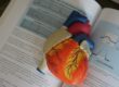

An echocardiogram uses sound waves to create moving images of your heart, and doctors rely on this test to study how the heart beats and pumps blood. When symptoms like chest pain or breathlessness appear, your physician may order one. Here is more information about this technology and how it can help diagnose heart conditions:
Measuring Pumping Strength
Ejection fraction measures how much blood your heart pushes out with each beat. An echocardiogram calculates this number directly. Because doctors need an accurate reading, they use the scan to track pumping strength over time.
Low numbers may signal heart failure or weakened muscle. The test flags these changes early. Doctors review the percentage, and they adjust treatment when values shift. Regular scans let your care team monitor progress.
Assessing Heart Function
Your heart moves constantly, and an echocardiogram captures that motion in real time. Doctors watch each chamber fill and empty. Since the images show live movement, physicians see how well the walls contract. This detail helps them understand your overall heart function.
The test shows the size and shape of your heart. If a chamber looks enlarged, that finding points toward specific conditions. The scan measures wall thickness, and it tracks blood flow through each section. Physicians compare these results against normal ranges.
The echocardiogram is a safe and noninvasive procedure commonly used in diagnosing heart conditions. It requires no special preparation, making it an accessible and straightforward test for most patients. While the process is simple, the detailed images it provides allow doctors to identify abnormalities and assess the severity of any underlying issues.
Identifying Valve Issues
Your heart has four valves that control blood flow between chambers. When a valve leaks or narrows, an echocardiogram reveals the problem. The scan shows how each valve opens and closes. Doctors measure the speed and direction of blood through them. Typical valve conditions include:
- Stenosis, where a valve becomes too narrow
- Regurgitation, where a valve allows blood to flow backward
- Prolapse, where a valve does not close properly
The test detects these issues without surgery. Since color flow imaging maps blood movement, physicians spot leaks quickly. They grade the severity, and they decide whether you need further care. This information guides treatment choices.
Spotting Muscle Damage
A heart attack leaves marks on the muscle tissue. An echocardiogram identifies areas that no longer move normally. When a section of muscle stops contracting, the scan highlights it clearly. Doctors use this to locate past or ongoing damage.
Damaged muscle typically appears thinner or scarred on the images. The test measures these weakened zones. Physicians review the affected regions, and they estimate how much function remains. This assessment shapes your recovery plan.
Some conditions the scan may reveal include:
- Reduced movement from a previous heart attack
- Thickened walls from long-term high blood pressure
- Fluid buildup around the heart
Since these findings appear on screen, doctors act on clear evidence. The scan supports faster, more accurate decisions.
Schedule an Echocardiogram Today
An echocardiogram gives your doctor a detailed view of your heart’s structure and function. Since it uses sound waves, the procedure carries no radiation risk. Contact your healthcare provider to ask whether an echocardiogram fits your needs. Bring a list of your symptoms and questions to the appointment, and your doctor will explain the results and outline any recommended steps.