
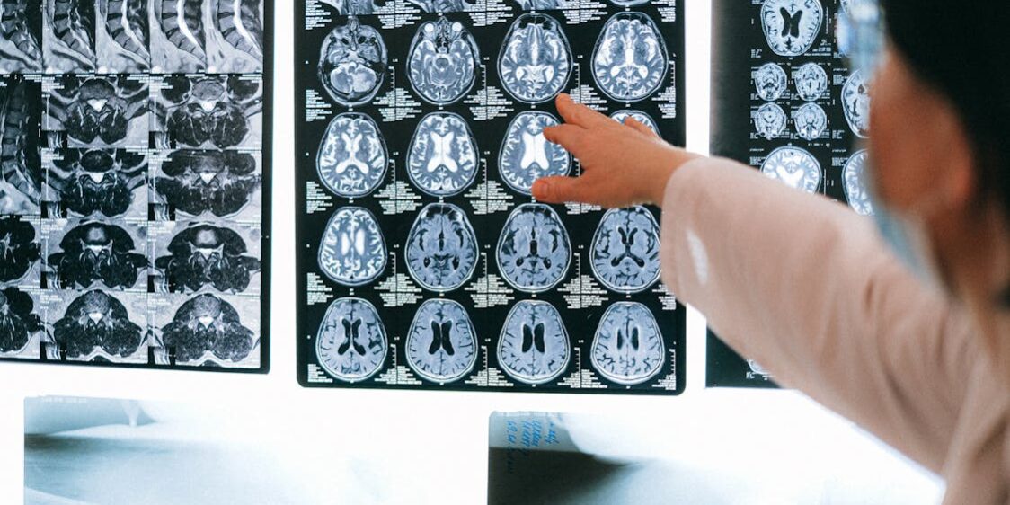
MRI technology is a valuable tool for examining the brain in greater detail without the need for invasive procedures. An MRI can help healthcare providers detect structural changes, evaluate neurological conditions, and support accurate diagnosis and treatment planning. Here is a closer look at some of the ways MRI technology is revolutionizing brain imaging:
Imaging Brain Structure
Modern MRI systems produce sharp and high-resolution images of the brain. This clarity allows imaging specialists to distinguish between different types of tissue with greater accuracy. White matter, gray matter, and fluid-filled spaces can now be mapped in fine detail, which was not possible with older technology. Higher-resolution imaging also means smaller structures can be captured more clearly; regions deep within the brain are now more accessible with advanced sequences. The improved detail supports thorough structural assessments without requiring invasive procedures.
Detecting Subtle Changes
One of the significant developments in MRI technology is its ability to detect small or early-stage changes in the brain. Functional MRI (fMRI) measures blood flow patterns, and it can highlight areas of activity during specific tasks. Diffusion tensor imaging (DTI) maps water diffusion along nerve fibers, revealing the organization and condition of white matter pathways. These techniques capture functional and connectivity data that adds another layer of detail to what a scan shows. Repeated scans over time also allow clinicians to track whether specific brain regions are changing, supporting monitoring in a structured and measurable way.
Supporting Diagnosis and Planning
Advanced capabilities provide valuable information that supports clinical decision-making. Here are some key areas where MRI plays a helpful role in diagnostic and planning workflows:
- Detailed anatomical mapping: This medical imaging tool produces precise images of brain structures, providing clinicians with a reference point for identifying regions of interest.
- Functional activity visualization: fMRI scans capture brain activity in real time, which is helpful when evaluating how specific areas respond to stimuli or tasks.
- Pre-procedural planning: Neurosurgeons and specialists use the data to plan procedures with greater spatial awareness.
- Monitoring over time: Sequential scans allow providers to compare images across visits, making it easier to track measurable changes in brain structure or function.
- Non-invasive tissue assessment: MRI provides detailed tissue information without the need for surgical access, a fundamental advantage for patient safety.
These capabilities make MRI a central tool in brain health workflows, supporting teams who need reliable and repeatable imaging data.
Making Scanning Comfortable
MRI does not use ionizing radiation, making it a safer option for repeated imaging than CT scans or X-rays. This is a key advantage when patients need multiple scans over time. The absence of radiation also makes MRI preferable when imaging younger patients or individuals with conditions that require frequent monitoring.
Comfort improvements have also advanced alongside the technology; newer MRI machines feature wider bore openings. This reduces the enclosed feeling that some patients find difficult to manage. Faster imaging sequences have shortened overall scan times, which lowers the time a patient spends lying still inside the machine. Noise-reduction systems and in-bore entertainment options are now standard in some modern facilities, contributing to a more manageable experience for patients.
Schedule Your MRI Appointment Today
MRI technology has changed brain imaging in measurable ways. These include clearer structural detail, improved detection of subtle changes, and stronger support for clinical planning. Each of these advances contributes to thorough and reliable brain imaging across a wide range of clinical scenarios. Contact a reputable medical imaging center in your area to schedule your MRI appointment today.