The human body relies on complex joint structures for basic mobility and balance, and at the foundation of this system lies the ankle. This joint facilitates walking, running, and navigating uneven terrain by connecting the lower leg directly to the foot. Learning about the underlying structure of this specific area helps individuals recognize signs of tissue damage. Because daily activities place continuous physical stress on the lower extremities, these structural components often face significant wear and tear over time. This puts the joint at risk of injury, such as ankle sprains.
What Is the Ankle’s Anatomy?
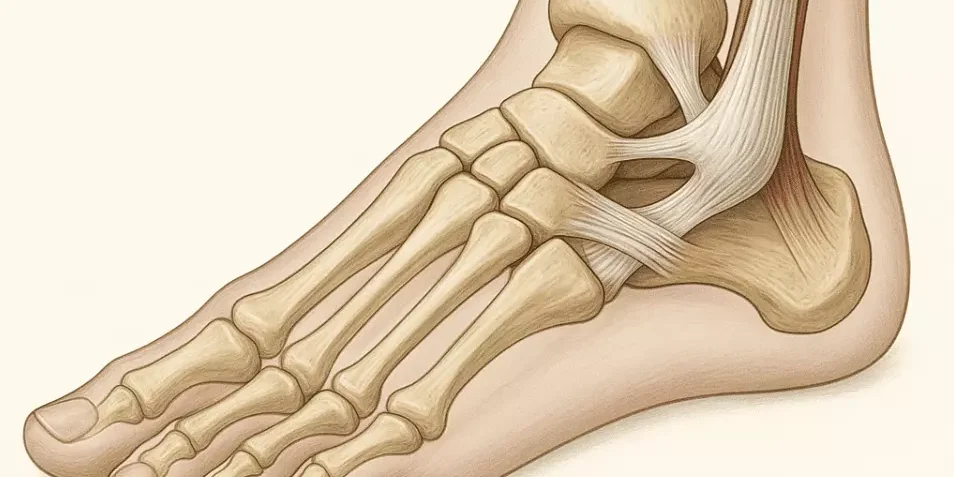
Classified as a synovial hinge joint, this anatomical structure allows the foot to move upward and downward. Three distinct bones form the main framework. The tibia and the fibula meet the talus bone located in the foot and fit together to create a stable cavity for movement. Soft tissues surround and support these bones. Cartilage coats the bone ends, providing a slippery, smooth surface that reduces friction during movement. This cartilage acts as a natural shock absorber for the skeletal system.
Ligaments serve as connective bands holding the bones securely in place. The medial ligaments anchor the inside, the lateral ligaments support the outside, and the syndesmotic ligaments bind the tibia and fibula together. Tendons connect the surrounding muscle groups to the bones, facilitating actions like pointing the toes or lifting the foot. A network of arteries supplies blood, and nerves relay electrical signals throughout the region.
What Is an Ankle Sprain?
Joint instability may lead to specific tissue damage known as an ankle sprain. This event occurs when a sudden movement forces the ligaments beyond their normal range of motion, causing the fibers to stretch or tear. Physicians classify these injuries into three distinct grades based on severity. A grade one classification involves minimal stretching of the ligament fibers without a complete tear. Grade two injuries signify a partial tear and typically result in moderate pain and swelling. A grade three classification indicates a complete rupture of the connective tissue, often leading to severe joint instability.
Symptoms generally manifest immediately following the injury event. Individuals may observe localized swelling, joint stiffness, and discoloration around the affected area. Putting weight on the affected limb can exacerbate the pain. Standard management typically involves resting the affected limb, applying ice packs, and using compression bandages to minimize swelling. Medical evaluation remains necessary to verify the exact grade of the sprain and to determine if additional treatments, such as immobilization or physical therapy, are required for full recovery.
What Other Conditions Affect It?
Beyond ligament damage, various other ailments impact this region of the lower extremities. Bone fractures represent a common issue and occur when excessive physical force breaks the tibia, fibula, or talus. Tendinitis affects the thick cords attaching muscle to bone. The Achilles tendon, located at the back of the heel, is particularly susceptible to inflammation and tearing due to overuse. Arthritis also impacts this joint and causes the protective cartilage to wear away over time. This deterioration results in chronic stiffness and discomfort during basic movements.
Seek Professional Podiatry Services
Proper diagnosis of lower extremity pain requires specialized medical knowledge. While minor soreness might resolve with brief rest periods, persistent swelling, severe pain, or an inability to bear weight indicates a need for immediate clinical evaluation. Addressing mobility issues promptly prevents chronic instability and long-term joint degradation. If symptoms persist or worsen after a traumatic event, schedule an appointment with a healthcare provider and discuss appropriate intervention strategies.